
|

|
Cancer Caught on Video
As chronicled in the NOVA program
"Cancer Warrior,"
one of Dr. Judah Folkman's most significant findings in a
career rife with discoveries was that cancerous tumors
appear to trigger the growth of new blood vessels, which
the tumors need to thrive. Here we present a series of
remarkable microscope views of various stages in cancer
growth and angiogenesis, or growth of new blood vessels.
Shot during experiments with laboratory chicken embryos
and mice, the clips follow a natural progression of cancer
spread, from early events up to the point when a tumor
requires angiogenesis to keep growing. The images, some
color and some black-and-white, were shot by Dr. Ann
Chambers and her colleagues at the University of Western
Ontario using a microscope outfitted with a video camera.
In many of the clips, you'll notice the camera focus
changing. Dr. Chambers wrote the captions that accompany
each clip.
Get video software:
QuickTime
|
RealVideo
|
|
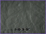
1. Early metastasis
View the clip in:
QuickTime
| RealVideo:
56K
|
ISDN+
This sequence shows an early step in the spread of a
cancer (a process called metastasis). A breast
cancer cell has traveled in the bloodstream and has
arrived at the liver, where it stops because it is
too big to keep moving through the tiny blood
vessels to get to another organ. The cell appears
bright because it has been labeled with a
fluorescent dye to help identify it.
|

|
|
|
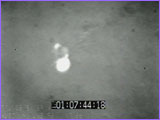
2. Escaping the bloodstream
QuickTime
| RealVideo:
56K
|
ISDN+
An early step in metastasis. This cancer cell has
escaped from the bloodstream and is partly wrapped
around the outside of a blood vessel. Because of
this, it does not need to attract new blood vessels
at this stage.
|
|
|
|
|
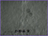
3. Cell division
QuickTime
| RealVideo:
56K
|
ISDN+
The first step in the growth of a new, metastatic
cancer. This cancer is made up of two cells, which
formed from the cell division of a single cell that
had escaped out of the bloodstream. It still does
not need angiogenesis at this stage, and it is
growing next to a pre-existing blood vessel.
|
|
|
|
|
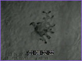
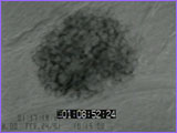
4. Small tumor
QuickTime
| RealVideo:
56K
|
ISDN+
This shows a very small metastatic cancer, early in
its development. This is a melanoma tumor, so it
appears black. It is growing around a blood vessel,
and you can see its three-dimensional shape as the
microscope focuses up and down through it. This
small tumor still does not need to attract new blood
vessels to support its growth, because the blood
vessel that it surrounds can support its growth at
this size.
|
|
|
|
|
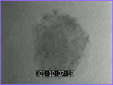
5. Attracting blood vessels
QuickTime
| RealVideo:
56K
|
ISDN+
As tumors grow larger, they begin to develop the
need for angiogenesis and must attract new blood
vessels if they are to keep growing. This small
melanoma cancer is beginning to show signs of blood
vessel activity inside it, and these might be
'angiogenic' new blood vessels.
|
|
|
|
|
6. New blood vessels (liver)
QuickTime
| RealVideo:
56K
|
ISDN+
This melanoma tumor is larger, about half a
millimeter wide, or roughly the size of a tiny grain
of sand. By this stage, the tumor needs to
continuously attract new vessels to keep on growing.
The normal liver tissue (lighter color) shows
normal, healthy blood flow, and the tumor (darker
color) shows new, angiogenic blood vessels with
irregular shapes and blood flow, especially visible
in the higher magnification clip.
|
|
|

|
|
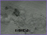
7. New blood vessels (body cavity)
QuickTime
| RealVideo:
56K
|
ISDN+
This melanoma tumor, also about a half a millimeter
wide, is growing on the body cavity wall of a mouse.
The black portion is the tumor and shows abnormal
'angiogenic' blood vessels, while the normal tissue
(lighter color) has more normal blood flow.
|
|
|
|
|
8. Continuous angiogenesis
QuickTime
| RealVideo:
56K
|
ISDN+
This tumor, about a tenth of a millimeter wide and
three-tenths of a millimeter long, is also growing
on the body cavity wall. It has attracted new blood
vessels to grow up to it from the normal muscle
tissue below. When tumors get to be this size, they
need continuous angiogenesis to keep on growing,
otherwise their growth will stop.
|
|
|
|
|
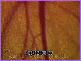
9. Normal, healthy blood vessels
QuickTime
| RealVideo:
56K
|
ISDN+
This view, taken with a color video camera, shows
normal, healthy blood vessels in mouse mammary
(breast) tissue. The red-filled vessels are blood
vessels, and the clear vessel (to the right of a
large blood vessel) is a lymph vessel. These blood
and lymph vessels show good flow and regular
branching patterns, typical of normal, healthy
organs.
|
|
|
|
|
10. Angiogenic blood vessels
QuickTime
| RealVideo:
56K
|
ISDN+
This view shows a breast tumor growing in mouse
breast tissue. The tumor appears green, because the
cancer cells were labeled with a fluorescent dye,
and the blood vessels appear black. These are new,
angiogenic vessels, and their structure and blood
flow look very irregular when compared to the
regular patterns seen in normal, healthy tissue (as
in the previous clip).
|
|
|
Note: video clips courtesy of Dr. Ann Chambers,
University of Western Ontario
Dr. Folkman Speaks
|
Cancer Caught on Video
Designing Clinical Trials
|
Accidental Discoveries
| How Cancer Grows
Help/Resources
|
Transcript
|
Site Map
|
Cancer Warrior Home
Editor's Picks
|
Previous Sites
|
Join Us/E-mail
|
TV/Web Schedule
About NOVA |
Teachers |
Site Map
|
Shop |
Jobs |
Search |
To print
PBS Online |
NOVA Online |
WGBH
©
| Updated February 2001
|
|
|