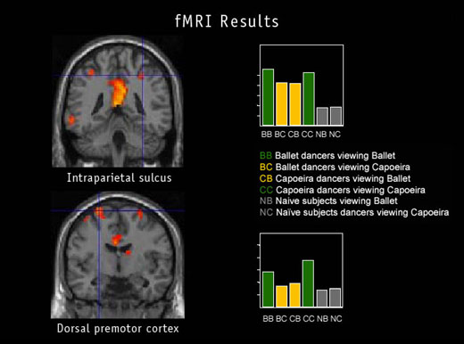
Daniel Glaser: What you are seeing on the left is two slices through the brain. We've got two sections there. The intraparietal sulcus, which is the upper section, is actually a little further back in the brain. That's the bit of the brain that is somewhere between the movement brain and the visual brain. It's, if you like, a visual-motor integration cortex. The area below it is further forwards in the head. That's the premotor, and that's the area of the brain that does complex motion planning, so that's the bit of the brain where we would expect to find representations of dance steps ready to be executed by the dancer when she was performing. Now, on the right you can see a couple of graphs. What we've done is to color-code in green the activity when you're seeing the thing that you can do and in yellow the activity when you're seeing the thing that you can't do. And you can see clearly that the two green bars are bigger than the two yellow bars in both cases. That's to say that the areas of brain are more activated when the two dancer groups are seeing what they can do than what they can't.
To the right of those colored bars, you can see two gray bars. That's people
like me and most of the people who will be looking at these images. This is
people whose brains and bodies are not trained to do either kind of movement.
And what you can see is that for those people the brain areas don't care
whether they are seeing ballet or capoeira. That means we've matched the
stimuli very well—they are using the same body parts—and your brain
is indifferent to those two if you're not an expert.
|
|||||||||||
|
|
|||||||||||
|
© | Created January 2005 |
|||||||||||