|

|

|
|
Windows on the Womb
Select any one of the techniques listed below to find out
more about it. The weeks during which doctors commonly use
such techniques are listed with each technique.
Enhanced Alpha Fetoprotein |
Amniocentesis |
Chorionic Villus Sampling |
Doppler device |
Fetal Echocardiography |
Ultrafast fetal MRI |
Nuchal Translucency Test |
Ultrasound scanning
Enhanced Alpha Fetoprotein or the Quad (AFP)
15 to 18 weeks
A protein produced by the baby's liver, alpha fetoprotein
(AFP) normally enters the mother's bloodstream. In this
test, blood drawn from the mother is examined for AFP; the
amount of AFP in her blood determines the level of risk for
disorders such as Down syndrome,
neural-tube defects,
abdominal-wall defects, and
Edwards syndrome. High levels may mean a
neural-tube defect or that some or all of the baby's brain
material is missing. A low level can be an indication of
Down syndrome. Because this is a screening test, showing
only the baby's level of risk, follow-up testing for an
abnormal level is recommended. There is no risk to the baby,
but because as many as 5 percent of all women test positive,
further testing often results. The vast majority of these
women turn out to carry healthy fetuses.
Amniocentesis
16 to 18 weeks
Obstetricians typically recommend the use of amniocentesis
for women more likely to be carrying a baby with
abnormalities, such as older women (35 and above) or women
with a family history of genetic diseases. An ultrasound
prior to the test determines the baby's location, and then a
specialist uses a small needle to withdraw about a
tablespoon of the amniotic fluid surrounding the baby. Cells
from the baby found floating in the fluid are cultured and
examined to look for chromosomal disorders. Doctors use the
test primarily to detect spina bifida or
Down syndrome, but also
Rh disease,
fetal anemia,
sickle-cell anemia, and to determine the
baby's sex. Late in pregnancy, doctors use amniocentesis to
find out if the baby's lungs are sufficiently developed and
thus able to withstand, if necessary, a medically required
premature birth. The U.S. Centers for Disease Control and
Prevention estimates that the rate of miscarriage following
amniocentesis is between one in 200 to 400 procedures.
Chorionic Villus Sampling (CVS)
10.5 to 13 weeks
As with amniocentesis, obstetricians may suggest using CVS,
short for chorionic villus sampling, to detect genetic
disorders such as Down Syndrome. In CVS,
specialists perform an ultrasound to determine the position
of the fetus and then remove fetal tissue by placing an
instrument through the cervix or abdomen. Unlike
amniocentesis, which can also detect Down syndrome, this
test can be carried out much earlier in pregnancy, and test
results are also available sooner. That means that if
parents decide to terminate a pregnancy based on the
results, they can do so as much as nine weeks earlier than
in the case of amniocentesis, creating fewer risks to the
mother's health. There is a 1 to 2 percent risk of
miscarriage following the procedure.
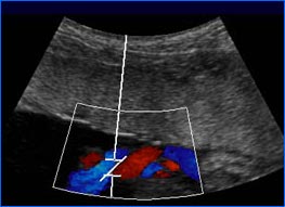
Doppler device
6 weeks to term
A Doppler device is a small, portable machine that uses
ultrasound waves to detect and magnify the baby's heartbeat.
Doctors use this test during most office visits to verify
that the baby is alive. After about the tenth week, a baby's
heart rate can vary between 120 and 170 beats per minute. In
the third trimester, obstetricians may use a variation of
this test, known as umbilical cord Doppler, to examine the
flow of nutrients between heartbeats, to ensure that the
baby is receiving adequate nourishment. There is no known
risk to the baby or the mother with this test.
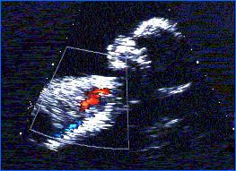
Fetal Echocardiography
14 weeks to term
This test is essentially a very detailed ultrasound
focussing on the structure and function of the heart.
Doctors use it only when either siblings or parents have a
history of heart defects, when other tests such as
amniocentesis have produced abnormal results, when the
mother has diseases that can affect the heart (such as
diabetes or
phenylketonuria), or when the fetus has
been exposed to certain drugs. Most experts conduct the test
between the 20th and 22nd week to
ensure that they can see the heart clearly. There is no
known risk to the baby or mother with this test.
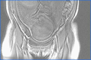
Ultrafast fetal MRI
second or third trimester
MRI (magnetic resonance imaging) relies on a magnetic field
and radio waves to "eavesdrop" on the body's electromagnetic
transmissions. An MRI image can clarify the diagnosis of a
fetal abnormality observed in an ultrasound and better
prepare parents and their doctors for any interventions that
may be needed to help the baby before or immediately after
birth. It is especially helpful for examining certain
tissues, such as the brain, that are encased in bone and
would be difficult or impossible to see using ultrasound.
MRI is not as widely available as ultrasound. It poses no
known risk to the baby or mother.
Nuchal Translucency Test (NT Scan)
11 to 14 weeks
This test uses ultrasound to examine the fold of skin on the
back of the baby's neck. At this early stage of development,
the skin is so thin that fluid accumulates between it and
the underlying structures. More fluid, which produces a
thicker fold, can be a sign that the baby has a chromosomal
abnormality, such as Down syndrome. This
test is available at a number of university medical centers
around the U.S. As with ultrasound, there is no known risk
to the baby or mother.
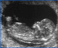
Ultrasound scanning
5 weeks to term
Many women will have at least one ultrasound during their
pregnancy. High frequency sound waves are directed at the
fetus and the returning "echoes" form a live-action picture
of the baby. Typically performed between 16 and 18 weeks, an
ultrasound provides a general check of the baby's anatomy
and can also help to date the pregnancy. Later on in
pregnancy, ultrasound can gauge the baby's growth and
development, determine the location of the placenta, and
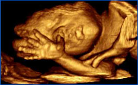
measure the amount of amniotic fluid. Three-dimensional
ultrasounds, which are now becoming available at some health
centers, provide a much clearer, more photographic image and
make it possible to observe the baby from any angle,
regardless of what position the baby is in during the
procedure. There is no known risk to the baby or mother.
Windows on the Womb—Glossary
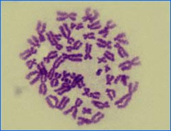
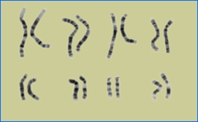
Down syndrome—In most cases caused by a third chromosome 21, Down
syndrome results in mental retardation and other
abnormalities. Children with Down syndrome have a widely
recognized characteristic appearance.
Neural-tube defects—An NTD occurs in the neural tube, the part of the
fetus that becomes the brain or spinal cord. NTDs result in
the partial or complete absence of the brain, or in an
opening of the spine. They are among the most common of all
serious birth defects.
Abdominal-wall defects—Abdominal-wall defects feature a soft bulge of
tissue or a small, localized swelling on the abdomen, most
often caused by a hernia. A hernia is an area where muscles
are weak enough to allow internal organs to protrude.
Edwards syndrome—Also known as trisomy 18, Edwards syndrome is
associated with a third chromosome 18, which causes multiple
physical abnormalities and severe mental retardation. Few
infants survive beyond their first year.
Rh disease—When the baby is Rh-positive and the mother is
Rh-negative, the mother's antibodies can cross the placenta
and attack the baby's red blood cells, resulting in
jaundice, anemia, brain damage, heart failure and death. Rh
disease occurs only when the mother has previously been
sensitized to Rh-positive red blood cells and has developed
antibodies to them.
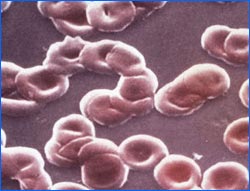
Fetal anemia—Fetal anemia occurs primarily when the mother's
blood type is incompatible with the baby's, leading to the
destruction of red blood cells in the baby's blood. This in
turn results in an oxygen deficiency for the baby.
Sickle-cell anemia—In this chronic inherited disease, the normally
round red blood cells become sickle- or crescent-shaped.
When these abnormally shaped cells move through small blood
vessels they can clog blood flow or break apart, causing
sudden severe pain in many areas of the body, damage, or
anemia.
Diabetes—Diabetes is a life-long disease in which the body
produces too little insulin or is unable to use the insulin
properly. The result can be dangerously high blood-sugar
levels, which, when untreated, starve cells of energy and
over time can damage the eyes, kidneys, nerves or heart.
Phenylketonuria—Phenylketonuria is a rare genetic disorder in which
the body is unable to properly metabolize the amino acid
phenylalanine, one of the eight essential amino acids found
in protein-containing foods. The accumulation of
phenylalanine in the blood and body tissues can cause severe
mental retardation and developmental delays if not
treated.
Watch the Program
|
The Stem-Cell Debate
|
Windows on the Womb
Great Expectations
|
How Cells Divide
|
How is Sex Determined?
Resources
|
Teacher's Guide
|
Transcript
|
Site Map
|
Life's Greatest Miracle Home
Search |
Site Map
|
Previously Featured
|
Schedule
|
Feedback |
Teachers |
Shop
Join Us/E-Mail
| About NOVA |
Editor's Picks
|
Watch NOVAs Online
|
To Print
PBS Online |
NOVA Online |
WGBH
©
| Updated February 2002
|
|
|
|